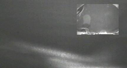
Wave Onset in Central Gray Matter – Original Image
The following figure
presents a sample original image got from the sequence captured during the
experiment for depression wave
visualization, presented with some enhancement on a prior
web page.
|
|
Besides the enhancement, the bottom part that does not pertain to the retina was labeled with a grayish color in order to give a better presentation and interpretation, as shown bellow. Then, the colored marks are added to set some references thus aiding to interpret the changing features of the wave onset and propagation.
|
|
|
|
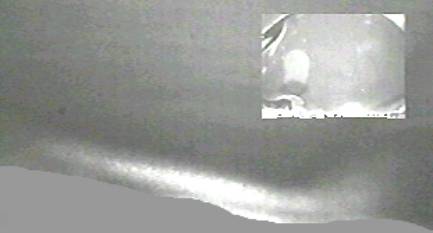
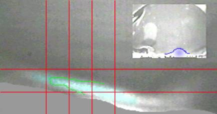
The anatomical structures present in the picture correspond to one octant of the chicken eye cut in a region near the pecten.
|
Spreading Depression |
|
|
|
LSI – Escola Politécnica –
University of São Paulo
and SENAC College of Computational Sciences
and Technology |
|
This page was created and is maintained by João E. Kögler
Jr.
Go to Homepage
|