Wave Onset in Central Gray Matter – Anatomical Structures
The following figures illustrate the anatomical structures and the experimental setup involved in the spreading depression studies in the retina conducted in the laboratory.
|
|
|
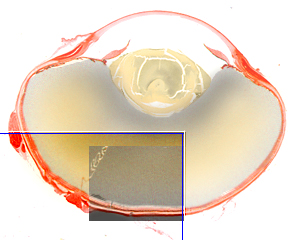
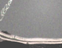
In the picture at the left it is depicted a cross-section of the chicken eye with a blue frame marking approximately one octant of the eye ball. Inside the octant , a rectangular window corresponds to the area captured with the camera, shown at left as an idealized image taken from an anatomical preparation. A sample actual image, as those presented at the wave onset web pages, is show below side by side with the above idealized image at approximately the same scale.
|
|
|
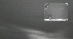
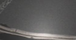
At the left is the actual image got from the experiment set and at the right is an adaptation of the above idealized image that was scaled, oriented and adjusted in contrast and brightness to display approximately the same conditions at the image at the right. These transforms were carried out just to the sake of didactic clarity.
|
Spreading Depression |
|
|
|
LSI – Escola Politécnica –
University of São Paulo
and SENAC College of Computational Sciences
and Technology |
|
This page was created and is maintained by João E. Kögler
Jr.
Go to Homepage
|