Intrinsic optical signals of lesions and penumbra zone
|
|
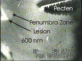
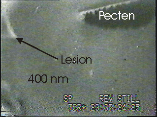
The two photos show the same scene illuminated with monochromatic light set at 600 and 400 nanometers respectively. In the experiment a mechanical lesion was made during vitreous removal. One can see a "fuzzy patch" of retina around the lesion under red illumination (600 nm); this patch is the so called Penumbra Zone. Note the absence of the intrisic optical signal in the penumbra zone, under blue illumination (400 nm).
 |
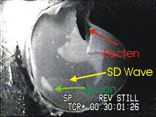 |
Thw two photos belong to the same experiment recorded with a black and white camera under white illumination. The gree arrow shows a patch of retina that suddenly becomes "fuzzy". Latter on a full blown lesion arises in the same region. The time interval between the two scenes is five minutes. Notice that in the central retina (around pecten) a big lesion was also developing, at the same time. The yellow arrow points to a retinal spreading depression wave bordering the small lesion.
This sudden onset of "fuzzy patches" similar to the one depicted in the photo was observed many times, sometimes concomitant with electrophysiological data. The abruptness of the onset suggests a phase transition in the system dynamics, heralded by the changes in the optical proprieties of the tissue.