Chicken Eye Anatomy
|
a. |
b. |
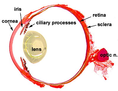
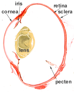
Figure: Eye anatomy for (a) mamals and (b) birds.
(Adapted from: http://www.class.cvm.uiuc.edu/j-eurell/eye14.htm)
The figure (a) depicts a tipical mamalian eye (it is from a dog). On figure (b) one can see an avian eye (from a chicken). They were represented on differing scales in order to show them as they would have the same size at the vertical diameter. At first we can poit the following differences between them:
- The avian eye is less spherical than the mamalian's. It is more crushed on the optical axis direction.
- The avian eye has the pecten structure which is said to have some rule on the retina nutrition and oxigenation. In the mammalian eye this is done by the choroid, which is a layer between the retina and the sclera. The avian eye is free from blood vessels, except at the pecten area.
This last property makes the chicken retina
a very interesting experimental substract. One can observe transport phenomena
across the retina without being disturbed by the network of blood circulation.
|
|
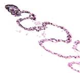
The
structure shown at the left is the so called pecten. |
|
Spreading
Depression |
|
|
|
LSI – Escola Politécnica –
University of São Paulo
and SENAC College of Computational Sciences
and Technology |
|
This page was created and is maintained by João E. Kögler
Jr.
Go to Homepage
|